Modeling the Maternal-Fetal Interface
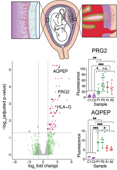
The maternal-fetal interface is essential for successful pregnancies as well as healthy postnatal and postpartum life. Formation of this interface begins with the implantation of an embryo into the uterine lining or endometrium, followed by development of the embryonically-derived placenta as the organ connecting mother and fetus. Deficiencies in any of the steps of this intricate process can lead to maternal and/or fetal mortality and morbidities, including conditions such as miscarriage, placenta previa and accreta, preeclampsia, and fetal growth restriction.
To model this intricate interface, I use a combination of mouse in vivo models and human in vitro organoid systems. I have developed one of the first mouse models of the impact of uterine injury on subsequent pregnancy outcomes. Since injury to the uterus from procedures such as C-section can lead to a host of pregnancy disorders, this mouse model brings us closer to a mechanistic understanding of how placenta previa, placenta accreta, infertility, and miscarriage can arise. In this animal model, uterine injury results in misspaced embryos (previa), thinned decidua with expanded invasive placental trophoblasts (accreta), fewer implanting embryos at the scar (infertility) and resorption (miscarriage). Intriguingly, embryo misspacing only occurs after injury during the diestrus but not estrus phase of the estrous cycle. Diestrus injuries lead to misexpression of COX/prostaglandin genes, thus pointing to a mechanism by which diestrus injury perturbs COX signaling long-term, resulting in defective uterine contractility and thus misspaced embryos in the subsequent pregnancy.


